Precise determination of camera intrinsic resolution, collimator spatial resolution, field size and linearity.
A means of lighting scintillation camera's crystal to determine response uniformity over the entire field.
Designed to simulate a patient’s neck.
Used to produce three 1 mm diameter parallel lines of tracer material spaced 7.5 cm apart.
Provides a multi-function simulation of the left ventricle, and can be used to evaluate SPECT imaging of cold defects within the "myocardium."
Provides the anatomically accurate three dimensional simulation of the radioisotope distribution found in the normal brain.
Designed for use in all circular and elliptical SPECT cylinders.
Consists of a body phantom, a fillable lung insert and an insert with six various size spheres that are fillable from the outside of the phantom.
Contains a fillable line source that runs parallel to the center axis of the cylinder and offset a distance of 4.5 cm.
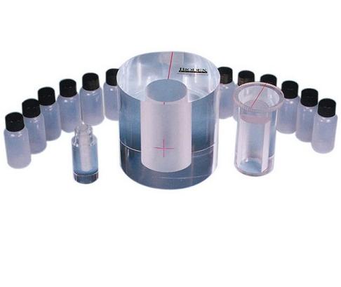
Consists of six concentric tubes that slide into each other. The innermost tube is fillable. The outer tubes are placed over the filled inner tube and imaged, adding a tube for each image.
Includes internal structures (three rods and six spheres) which, when imaged with both modalities, can demonstrate how accurately the two image sets are aligned.
Provides consistent performance information for any SPECT or PET system.
The flangeless SPECT Phantom meets the requirements set by ACR.
Used for the evaluation of non-uniform attenuation and scatter compensation methods. The phantom consists of a large, body-shaped cylinder with lung, liver and spine inserts.
The Lung-Spine Phantom consists of two chambers that are shaped to simulate the lungs.